

This service includes tissue preparation, sectioning, immunostaining, mounting, coverslipping and labeling the slides. As a result, you will receive up to 60 immunostained sections per brain or per tissue block ready for microscopic observations.
Procedure: Following cryoprotection, tissue will be rapidly frozen in isopentane pre-cooled to -70°C. The frozen tissue will then be cut on a cryostat and collected in our unique section cryopretection solution. Subsequently, sections cut from various levels (or the levels of your choice) will be processed free-floating for immunohistochemistry with one specific antibody according to the avidin-biotin-complex (ABC) method¹ (cf. photo samples below).
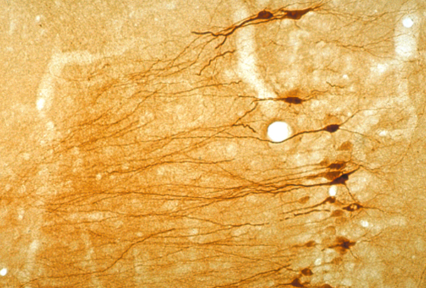 |
Heat shock protein-immunoreactivity. This 30 µm cryostat section was cut from the dorsal hippocampus of a rat that survived for 15 hrs after the injection of aminooxyacetic acid into the entorhinal cortex (for details, cf. Neurosci. Lett. 147:185-188, 1992). The section was processed free-floating according to avidin-biotin-complex method (click to see enlarged photo). |
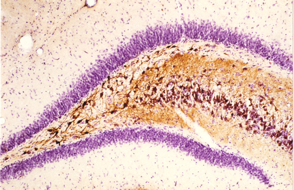 |
Heat shock protein-immunostained section counterstained with thionin. This 30 µm cryostat section was cut from the dorsal hippocampus of a rat that survived for 15 hrs after the injection of aminooxyacetic acid into the entorhinal cortex (for details, cf. Neurosci. Lett. 147:185-188, 1992). The section was processed for heat shock protein-immunostaining (brown) and then counterstained with thionin (click to see enlarged photo). |
 |
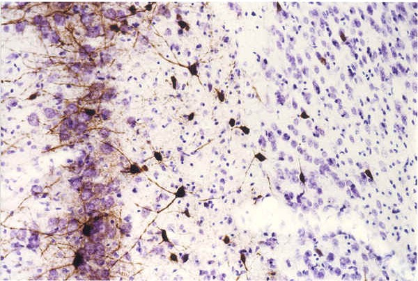
OX18-immunostained section counterstained with thionin. This 30 µm cryostat section was cut from the entorhinal cortex of a rat that survived for 24 hrs after the injection of aminooxyacetic acid into the entorhinal cortex. The section was processed for OX-immunostaining and then counterstained with thionin. Note the expression of OX-18 immunoreactivity (brown) in activated microglia within layer III (click to see enlarged photo). |
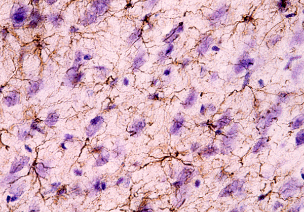 |
GFAP-immunostained section counterstained with thionin. This 30 µm cryostat section was cut from the entorhinal cortex of a normal rat. The section was processed for GFAP-immunostaining and then counterstained with FD thionin solution (cf. Products, Cat. #PS101). Note GFAP-immunoreactivity (brown) in the processes of astrocytes (click to see enlarged photo). |
 |
Parvalbumin-immunostained section counterstained with thionin. 30 µm cryostat section through the medial entorhinal cortex of a rat that survived for 24 hrs after kainic acid administration, showing the preferential loss of neurons in layer III and relative resistance of parvalbumin neurons (for details, cf. J. Neurosci. 15:6301-6313, 1995) (click to see enlarged photo). |
Remarks:
Reference: