

Counterstain is often used to reveal cells that are not labeled by the primary staining. For example, tissue sections that have been previously processed for immunohistochemistry or other labelings (e.g. TUNEL, ISNT) may be counterstained with histological dyes such as thionin (blue), neutral red (red), or methyl green (green) (cf. photo samples below).
SG503-C Counterstain with cresyl violet (cf. Products, Cat. #PS102)
SG503-E Counterstain with eosin Y (cf. Products, Cat. #PS103)
SG503-H Counterstain with hematoxylin (cf. Products, Cat. #PS104)
SG503-M Counterstain with methyl green (cf. Products, Cat. #PS105)
SG503-R Counterstain with neutral red (cf. Products, Cat. #PS106)
SG503-T Counterstain with thionin (cf. Products, Cat. #PS101)
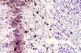 |
Parvalbumin-immunostained section counterstained with thionin. 30 µm cryostat section through the medial entorhinal cortex of a rat that survived for 24 hrs after kainic acid administration, showing the preferential loss of neurons in layer III and relative resistance of parvalbumin neurons (for details, cf. J. Neurosci. 15:6301-6313, 1995). |
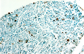 |
TUNEL-labeled section counterstained with methyl green. This 10 µm paraffin section was cut from a dorsal root ganglion of a mouse embryo (E17). The section was processed for detecting neuronal apoptosis (brown) with FD NeuroApop™ Kit (cf. Products, Cat. #PK201) and then counterstained with methyl green. |
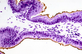 |
Cytokeratin 18-immunostained section counterstained with cresyl violet. This 12 µm frozen section of the rat prostate was processed for Cytokeratin 18-immunoreactivity (brown) and was then counterstained with FD cresyl violet solution (cf. Products, Cat. #PS102). |
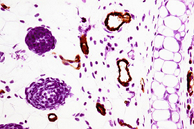 |
CD31-immunostained section counterstained with hematoxylin. This 7 µm paraffin section of the mouse ear was processed for CD31-immunoreactivity (brown) and was then counterstained with FD hematoxylin solution (cf. Products, Cat. #PS104). |
Remark: