FD NeuroApop™ Kit
FD NeuroApop™ Kit is specifically designed for the detection of neuronal apoptosis in tissue sections from the central nervous system based on the principle of in situ DNA nick-end labeling (TUNEL) technique¹. The assay uses terminal deoxynucleotidyl transferase to catalyze the incorporation of biotinylated deoxyuridines onto the free 3′-hydroxyl termini of DNA fragments, which are considered one of the most characteristic features of apoptosis2, 3. The integrated biotins are amplified and visualized with the avidin-biotin-complex (ABC) method4, enabling light microscopic identification.
The reagents and procedure of FD NeuroApop™ Kit have been optimized to achieve a high degree of both specificity and sensitivity for detecting apoptotic neurons with the lowest background. This kit can be used with frozen and paraffin sections, as well as cultured cells (cf. photo samples below). The procedure of the kit takes approximately 4 hours.
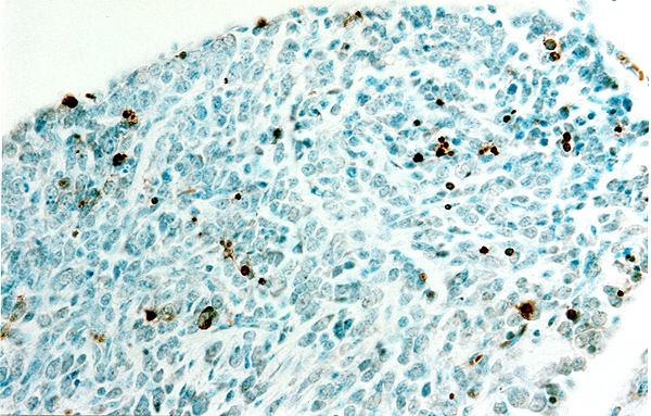 |
Detection of apoptotic neurons with FD NeuroApop™ Kit.
Paraffin section (10µm) cut from a dorsal ganglion of a mouse embryo (E17). The section was processed with FD NeuroApop™ kit and counterstained with methyl green. Sections courtesy of Drs. Michael Vogel and Lisa Qiu (click to see enlarged photo). |
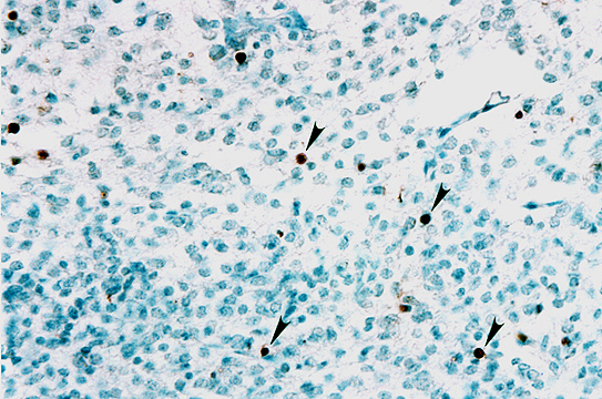 |
Detection of apoptotic neurons with FD NeuroApop™ Kit.
Paraffin section (10µm) cut from the inferior colliculus of a mouse embryo (E17). The section was processed with FD NeuroApop™ kit and counterstained with methyl green. Sections courtesy of Drs. Michael Vogel and Lisa Qiu (click to see enlarged photo). |
 |
Detection of apoptotic neurons in a rat model of stroke.
20 µm cryostat section was cut from the rat striatum of a stroke model. The section was processed for detecting neuronal apoptosis with FD NeuroApop™ Kit and then counterstained with FD methyl green (click to see enlarged photo). |
Kit contents:
Part I (Store at -20°C)
- Digestive Enzyme 2 ml x 4
- Reaction Solution A 2 ml x 2
- Reaction Solution B 85 µl
- Reaction Solution C 60 µl
- Chromogen Solution 20 ml
Part II (Store at 4°C)
- Equilibration Buffer 20 ml
- Detection Reagent 5 ml
- 10x Phosphate-Buffered Saline 250 ml x 2
Materials required but not included:
- Double distilled water
- Humidified chamber
- Incubator or waterbath (30°C)
- Histological supplies and equipment, including microscope slides, glass coverslips, staining jars, fine-tipped forceps, ethanol, xylenes or xylene-substitutes, mounting medium, and a light microscope.
References:
- Gavrieli Y, Sherman Y, and Ben-Sasson SA. (1992) Identification of programmed cell death in situ via specific labeling of nuclear DNA fragmentation. J. Cell Biol. 119:493-501.
- Wyllie AH. (1980) Glucocorticoid-induced thymocyte apoptosis is associated with endogenous endonuclease activation. Nature. 284:555-6.
- Arends MJ, Morris RG, and Wyllie AH. (1990) Apoptosis: the role of the endonuclease. Amer. J. Pathol. 136:593-608.
- Hsu SM, Raine L, and Fanger H. (1981) Use of avidin-biotin-peroxidase complex (ABC) in immunoperoxidase techniques: a comparison between ABC and unlabeled antibody (PAP) procedures. J. Histochem. Cytochem. 29:577-80.
|