

The combination of hematoxylin and eosin stains (H & E, cf. Products, Cat. #PS104 & PS103) is one of the most widely used staining techniques in histology and histopathology. Hematoxylin imparts a blue color to the basophilic tissue elements including nuclear chromatin, while eosin gives various shades of red color to acidophilic tissue components such as collagen, cytosol, musculature and erythrocytes (cf. photo samples below).
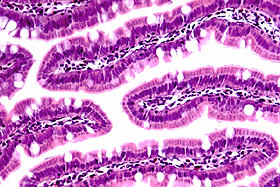 |
H&E stain. This 5 µm paraffin section of rat intestine was stained with FD hematoxylin (blue) and FD eosin Y (red). |
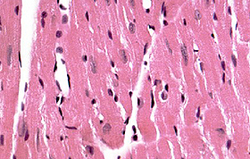 |
H&E stain. This 5 µm paraffin section of mouse heart was stained with FD hematoxylin (blue) and FD eosin Y (red). |
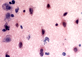 |
H&E stain. This 20 µm cryostat section from the rat cortex of a stroke model was stained with FD hematoxylin (blue) and FD eosin Y (red). Note that damaged neurons are stained red in the cytoplasm and most of them have a condensed, irregular-shape and darkly stained nucleus. |
Remark: