

This service includes tissue preparation, sectioning, immunostaining, mounting, coverslipping and labeling the slides. As a result, you will receive up to 60 immunostained sections per brain or per tissue block ready for microscopic observations.
Procedure: Following cryoprotection, tissue will be rapidly frozen in isopentane pre-cooled to -70°C. The frozen tissue will then be cut on a cryostat and collected in our unique section cryoprotection solution (cf. Products, Cat. #PC101). Subsequently, sections cut from various levels (or the levels of your choice) will be processed free-floating for immunostaining with 1 specific antibody according to the indirect immunofluorescence method¹ (cf. photo samples below).
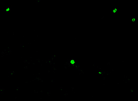 |
Cofocal image of BrdU-immunoreactivity. 30 µm cryostat section was cut from the hippocampal dentate gyrus of a mouse that survived for 24 hrs after the injection with 5-bromo-2-deoxyuridine (BrdU). This section was processed free-floating according to the indirect fluorescence method. |
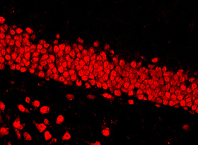 |
Cofocal image of NeuN-immunoreactivity. The same section as shown above was processed free-floating for NeuN-immuoreactivity according to the indirect fluorescence method. Note NeuN-labeled granule cells and polymorphic neurons in the dentate gyrus. |
 |
Colocalization of BrdU- and NeuN- immunoreactivities. A digital overlay of the 2 images shown above. Note that the regions of colocalization, reflecting the additive effect of superimposed green and red pixels, appear in yellow. |
Remarks:
Reference: