

This service includes tissue preparation, sectioning, immunostaining, coverslipping and labeling the slides. As a result, you will receive up to 60 immunostained sections per brain or per tissue block ready for microscopic observations.
Procedure: Following cryoprotection, tissue will be rapidly frozen in isopentane pre-cooled to -70°C. The frozen tissue will then be cut on a cryostat and mounted on gelatin-coated microscope slides (cf. Products, Cat. #PO101). Subsequently, sections cut from various levels (or the levels of your choice) will be processed on slides for immunostaining with one specific antibody according to the indirect immunofluorescence method¹ (cf. photo samples below).
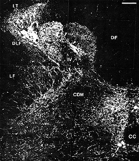 |
Substance P-immunofluorescence in the spinal cord. 10 µm cryostat section was cut transversely from the chicken spinal cord. This section was processed on slide according to the indirect fluorescence method (for details, cf. J. Comp. Neurol. 278:253-264, 1988). Note substance P containing fibers mainly in the dorsolateral funiculus, Lissauer’s tract and the dorsal horn. |
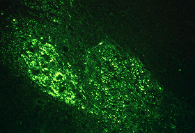 |
Substance P-immunofluorescence in the spinal cord. A 10 µm cryostat section of the chicken spinal cord was processed as described above. Note dense substance P immunoreactivity in the dorsolateral funiculus and the dorsal horn. |
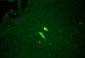 |
Vasoactive intestinal polypeptide-immunofluo-rescence in the spinal cord. A 10 µm cryostat section of the chicken spinal cord was processed on slide according to the indirect fluorescence method. Note 2 large neurons containing vasoactive intestinal polypeptide in the nucleus of the dorsolateral funiculus (for details, cf. J. Comp. Neurol. 278:253-264, 1988). |
Remarks:
Reference: