

This service includes tissue preparation, sectioning, silver-staining, mounting, coverslipping and labeling of slides. As a result, you will receive up to 40 silver-stained sections per brain or per tissue block ready for microscopic observations.
Procedure: Following cryoprotection, tissue will be rapidly frozen in isopentane pre-cooled to -70°C. The frozen tissue will then be cut on a cryostat and collected in our unique section cryoprotection solution (cf. Products, Cat. #PC101). Subsequently, sections cut through various levels (or the levels of your choice) will be processed free-floating for the detection of neurons undergoing degeneration with Gallyas silver staining technique¹.
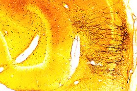
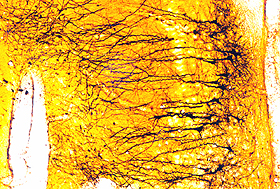
This technique was originally described by Gallyas et al.¹ and later modified². It is particularly useful for the detection of early neuronal injury in the central nervous system of experimental animals. Typically, both the neuronal perikarya and processes are silver-stained. This technique permits the morphological categorization of damaged neurons and the detection of subtle changes in the morphology of cell bodies and processes (cf. photo samples below).
 |
 |
Remarks:
References: