

This service includes sectioning, ISNT labeling, coverslipping and labeling of slides. As a result, you will receive up to 40 ISNT-labeled sections per brain or per tissue block ready for microscopic observations.
Procedure: Unfixed frozen tissue will be cut on a cryostat and mounted on proteinase-resistant microscope slides (cf. Products, Cat. #PO102). Sections cut through various levels (or the levels of your choice) will be processed on slides for the detection of neurodegeneration with in situ nick translation technique¹.
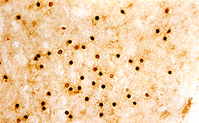
This technique, originally described by Gold et al. (1993)¹, labels the nuclei of neurons undergoing DNA fragmentation. This method is based on the ability of DNA polymerase I to catalyze the polymerization of deoxyuridines onto the ends of DNA fragments. The integrated biotins are amplified and visualized by the avidin-biotin-complex methods² (cf. photo sample below).
Remarks:
References: