

Timm’s sulfide silver staining has been used to visualize a variety of metals in brains and other tissues¹. Among these are the trace metals essential for life, such as Zn, Cu, Fe, Co, and Ni, as well as toxic metals, e.g., Hg, Cd, Pb, As, Bi, TI, Au and Ag. This method, originally developed by Timm², was later modified3. The principal of the technique is based on sulphide-precipitation of metals in tissue followed by a physical development. During the latter stage, the metal sulphides catalyze the reduction of silver ions by reducing agents. This technique has proven to be particularly useful in visualizing zinc-containing neurons and the detection of newly sprouted axons and axon terminals within the central nervous system (cf. photo samples below).
This service includes tissue preparation, sectioning, staining, coverslipping and labeling the slides. As a result, your will receive up to 40 Timm’s sulfide silver stained sections per brain or per tissue block ready for microscopic observations. A set of Nissl (or H&E) stained sections adjacent to those used for silver-staining can also be provided at a low cost.
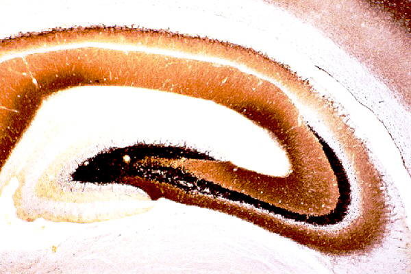 |
Timm’s staining of the rat hippocampus. 30 µm cryostat section cut through the dorsal hippocampus of a normal rat was processed for Timm’s histochemistry. Note that the highest density of reaction product is present in mossy fibers traveling in the polymorphic layer of the dentate gyrus and the stratum lucidum of CA3. |
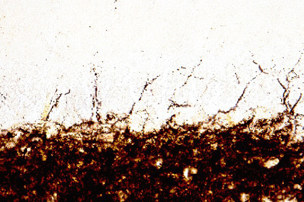 |
Timm’s staining in the dentate gyrus of a normal rat. High magnification of the polymorphic, granule cell and molecular layers of the dentate gyrus from the same section as shown above. Note little reaction deposits in both the granule cell and molecular layers. |
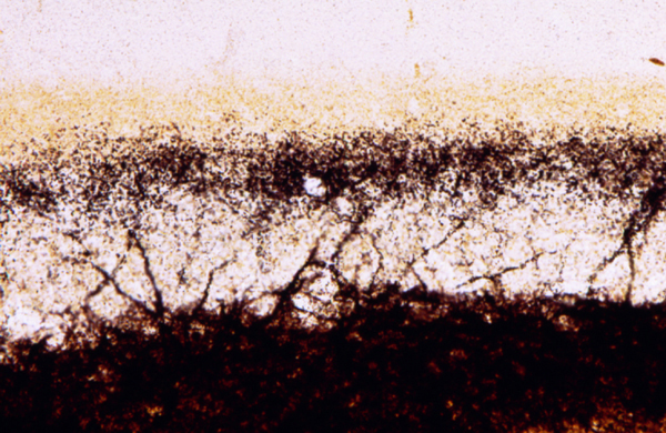 |
Timm’s staining in the dentate gyrus of a rat model for epilepsy. High magnification of the dentate gyrus from a rat that had developed chronic seizures. Note a dense plexus of reaction product in the inner (supragranular) layer of the dentate gyrus (compare with the photo above). |
Remarks:
References: