PK301 and PK301A: Degenerating neuronal somata, axons, and terminals in both the brain and the spinal cord

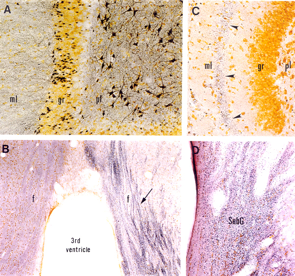
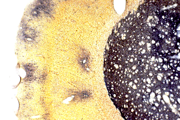
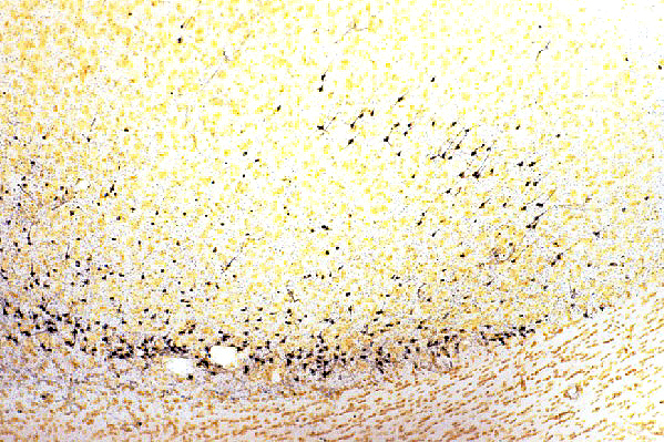
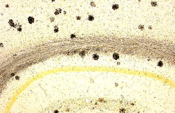
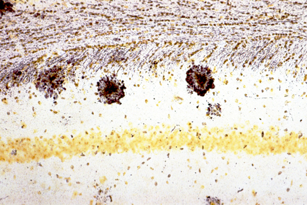
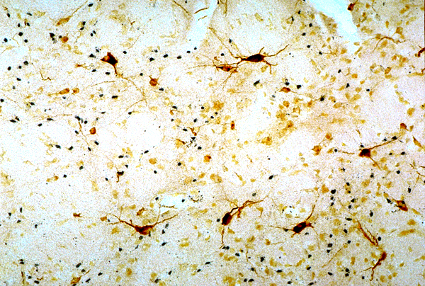
Detection of neurodegeneration with FD NeuroSilver™ Kit I.  A: Section (40 µm) through the dentate gyrus of the hippocampus from a rat injected with kainic acid (10 mg/kg, s.c.), showing degenerating neurons and processes (black) in the polymorphic layer (pl) and the molecular layer (ml), respectively.  B: Coronal section (30 µm) through the septum of a rat, killed at 10 days following a unilateral transection of the fimbria. Note degenerating axons (indicated by the arrow) in the ipsilateral fornix.  C: Horizontal section (40 µm) through the hippocampal dentate gyrus of a rat killed at 5 days after the intra-entorhinal injection with aminooxyacetic acid. Note numerous degenerating axon terminals (arrowheads) in the middle zone of the molecular layer (ml), the terminal field of entorhinal neurons killed by the drug injection (for details, cf. Neuroscience 82:1165, 1998).  D: Coronal section (40 µm) throught the lateral geniculate nucleus of a rat killed at 5 days after a unilateral visual cortex aspiration. Note dense degenerating axon terminals in the subgeniculate nucleus (SubG). Section courtesy of Dr. E.-Y. Chen, Rush University (D).

Detection of neurodegeneration in the rat brain of a stroke model.  40 µm cryostat section was cut coronally through the striatum of a rat used as an animal model of stroke. The section was processed for demonstrating neuronal damage with FD NeuroSilver™ Kit I. Note the accumulation of metallic silver grains (black) in both the striatum and the cortex, indicating the presence of neurodegeneration.

Detection of neurodegeneration in the rat brain of an epilepsy model.  40 µm cryostat section through the cortex of a rat used as an animal model of epilepsy. Note degenerating neurons (black) in the deep layers.

PK301 and PK301A: Detection of amyloid plaques in the brain of transgenic mice

Detection of neurodegeneration and amyloid plaques in the mouse brain of a model for Alzeimer’s disease.  40 µm cryostat section was cut coronally through the dorsal hippocampus of a transgenic mouse used as a model for studing Alzeimer’s disease. The section was processed for detecting both neuronal damage and amyloid plaques with FD NeuroSilver™ Kit I. Note silver-stained plaques in both the hippocampus and the cortex.
High mignification of the hippocampal CA1 area as shown above. Note that in addition to silver-stained amyloid plaques, numerous degenerating fibers are also present in the corpus callosum.

PK301 and PK301A: Neurodegeneration and/or amyloid plaques in tissue sections that have been processed for immunohistochemistry

Detection of neurodegeneration in the rat brain of a model for Huntington’s disease.  40 µm cryostat section was cut coronally through the striatum of a rat that received a focal injection of quinolinic acid. The section was processed first for bcl2-immunoreactivity and then for detecting neuronal damage with FD NeuroSilver™ Kit I. Note that degenerating neurons (black) intermingle with survived bcl2-positive neurons.